INTRODUCING
Bobade Accident Hospital
Bobade Accident Hospital, established in 1988, is a premier orthopedic and trauma care center located in Baramati, Maharashtra. Strategically situated near the central bus stand, the hospital caters to a population exceeding 2 lakh, including underserved rural regions. Over the decades, it has evolved into a referral center for advanced orthopedic, arthroscopic, and spine interventions.
Bobade Accident Hospital has evolved into a leading healthcare facility, renowned for delivering the highest quality accident and emergency services.
MORE ABOUTOUR SERVICES
Special High-quality Services
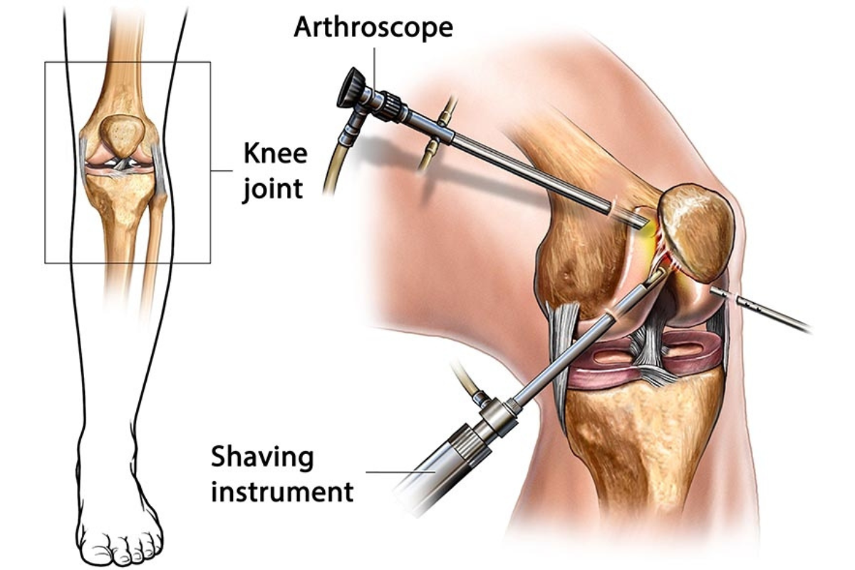
We offer advanced arthroscopy surgery performed by expert orthopedic surgeons, ensuring minimally invasive treatment for joint conditions with faster recovery and minimal discomfort.
READ MORE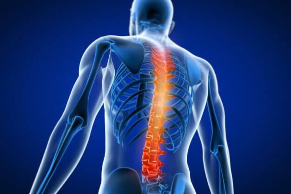
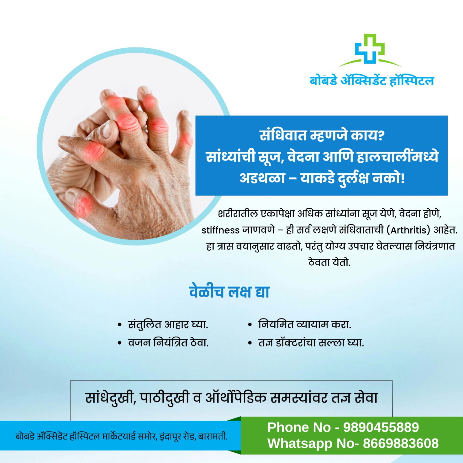
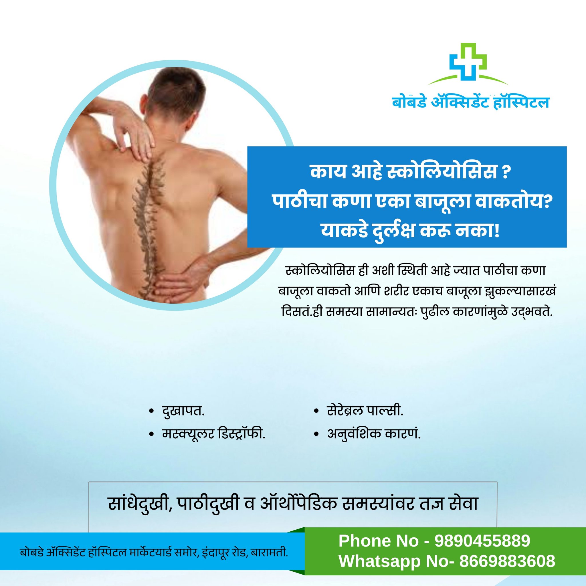
Our specialized spine surgery services provide effective treatment for spinal disorders, using advanced techniques to relieve pain, restore mobility, and improve quality of life
READ MORE
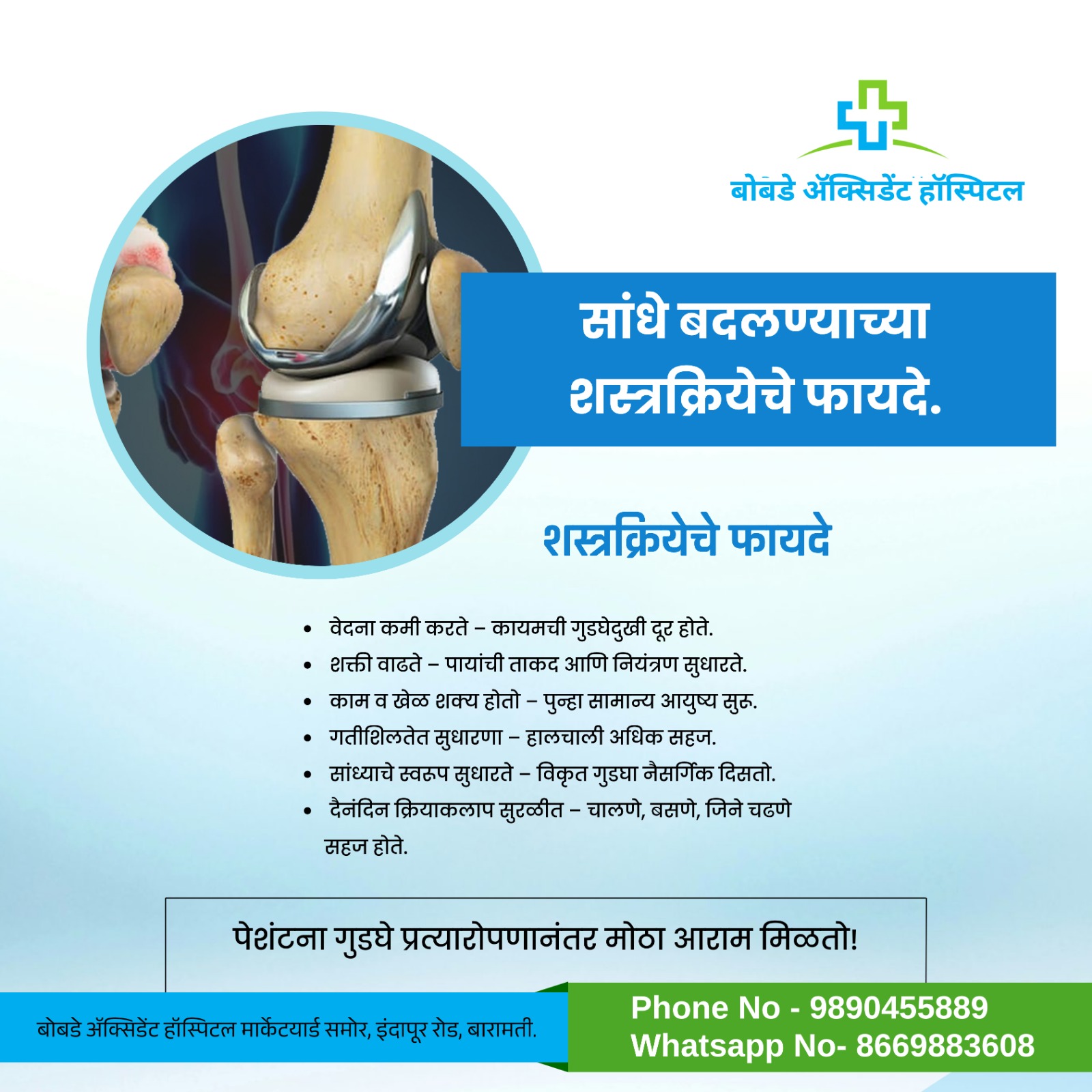
We offer expert joint replacement surgery using the latest technology to restore mobility, reduce pain, and help patients return to an active lifestyle.
READ MOREMEET OUR EXPERIENCED TEAM
Our Dedicated Doctors Team

AWESOME TIPS
Our Latest Health Tips


HAPPY CLIENTS
What Our Patients Are Saying

Life-saving care when it mattered most!“I was brought to Bobade Accident Hospital after a serious road accident. The emergency team responded immediately, and the doctors stabilized my condition within minutes. From surgery to physiotherapy, every department worked with care and efficiency. I am truly thankful to the entire staff for giving me a second chance at life.
Rahul Patil
Expert treatment with a personal touch.My mother was admitted here with a broken hip after a fall. The orthopedic team at Bobade Accident Hospital performed the surgery with great skill, and her recovery was closely monitored by attentive nursing staff. We were comforted not just by the treatment, but by the warmth and care shown throughout her stay.
Sneha Deshmukh
Quick diagnosis, trusted resultsI visited the hospital for sudden chest pain. Within minutes, the doctors had conducted X-rays, ECG, and blood tests — everything was available under one roof. The efficiency of the staff and the availability of emergency diagnostics truly sets this hospital apart.
Imran Shaikh
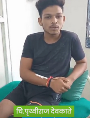
VIDEO TESTIMONIAL
What Patients Says About Us